1. Describe the main finding.
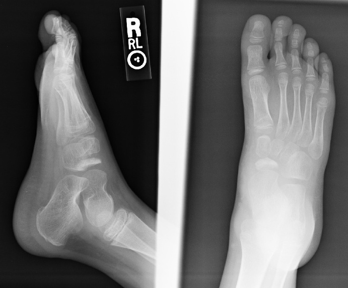
The navicular appears compressed and sclerotic.
2. Do you have a diagnosis? a differential diagnosis?
This is the classic radiographic presentation for Kohler's disease. Differential diagnosis includes normal irregular ossification of the navicular. Fragmentation is not always present but helps to confirm the diagnosis.
3. What is the etiology of the pertinent finding?
Kohler's disease most commonly occurs between 3-7 years of age and is much more common in males. It is the result of avascular necrosis however the exact cause precipitating the loss of blood supply is not always clear. Some but not all cases may be due to trauma, others may be due to altered compressive forces around the calcaneus which may affect the blood supply.
Classic presentation is pain, tenderness, and possible limping. Physical evaluation confirms local pain in the region of the navicular, swelling, and decreased range of motion. These findings will help differentiate the diagnosis from a normal developmental variation. Approximately 1/3 of cases will present with a history of trauma. Up to 25% of cases will have a bilateral presentation. Rest, restricted activity, and in some cases arch supports may be prescribed. Casting for several weeks may be performed if there is severe pain or if the condition worsens. Symptoms may require up to 2 years to completely resolve without long term effects.