 |
 |
 |
 |
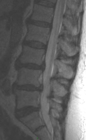
1. Aside from disc changes and spondylosis, what additional finding is present.
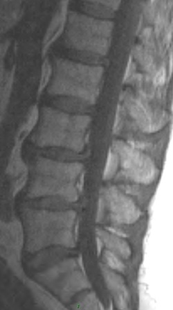
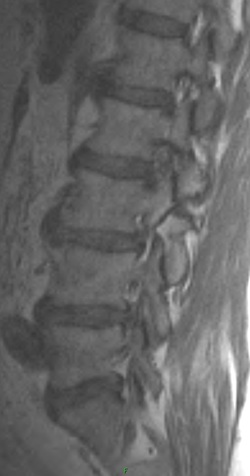
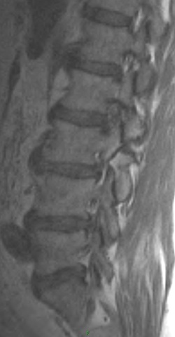
There is evidence of a grade 1 anterolisthesis of L3 on L4. The second set of images show non acute bilateral pars fractures at L3, responsible for the anterolisthesis.
2. What are the types of spondylolisthesis? Pertaining to the anterolisthesis, which type does the patient have?
The patient has an isthmic spondylolisthesis of L3. This is a unique case in that L5 is by far the most common location for this type of spondylolisthesis, with L4 less common, and L3 even more uncommon.
Five main types of spondylolisthesis are identified.
(1) Dysplastic - this involves congenital dysplasia of the posterior elements producing ineffective posterior stabilization. Normal weight bearing produces slippage of the vertebral body.
(2) Isthmic - 3 main subtypes exist -
a) lytic (spondylolysis) - most common type among young patients, may represent a stress fracture of the pars, most commonly involves L%
b) elongated but intact pars - thought to be repeated microfractures or fractures of the pars that have healed over time, occurs at L5
c) acute pars fractures - rare, cause by severe acute trauma
(3) Degenerative - most common type overall, most common over 50 years of age, much more common in women than men, slippage is usually 25% or less, more likely to produce spinal stenosis
(4) Post traumatic - rare, fracture in a supporting structure not involving the pars
(5) Pathologic - due to localized or generalized bone disease involving the pedicle, pars, superior, and/or inferior facets Plain films are usually diagnostic. In cases where developing spondylolysis is suspected, SPECT or MRI may be useful to further evaluate the pars. Spondylolisthesis is most commonly graded based on the percent of slippage in relation to the length of the verterbral endplate:
Anterior and posterior spondylolisthesis is best visualized and measured on lateral radiographs of the spine. In the lumbar spine oblique views may be performed to better visualize the pars region in cases of isthmic type spondylolisthesis.
3. What is the appropriate way to asses stability?
Lateral flexion and extension radiographs or lateral neutral views with traction and compression can be performed to assess stability. A change of 4 mm in the anterolisthesis in either flexion or extension is suggestive of instability.
4. What are treatment options for spondylolisthesis?
Conservative care is indicated for stable anterolisthesis. This may include any or all of the following - chiropractic care, bracing, exercises to strengthen abdominal and back musculature, analgesics, and restriction of physical activity in cases of acute symptoms. An unstable spondylolisthesis is more likely to be symptomatic and tends to have a poorer prognosis with conservative treatment. Unstable anterolisthesis may be treated with posterior lumbar intervertebral fusion.
There is evidence of a grade 1 anterolisthesis of L3 on L4. The second set of images show non acute bilateral pars fractures at L3, responsible for the anterolisthesis.
2. What are the types of spondylolisthesis? Pertaining to the anterolisthesis, which type does the patient have?
The patient has an isthmic spondylolisthesis of L3. This is a unique case in that L5 is by far the most common location for this type of spondylolisthesis, with L4 less common, and L3 even more uncommon.
Five main types of spondylolisthesis are identified.
(1) Dysplastic - this involves congenital dysplasia of the posterior elements producing ineffective posterior stabilization. Normal weight bearing produces slippage of the vertebral body.
(2) Isthmic - 3 main subtypes exist -
a) lytic (spondylolysis) - most common type among young patients, may represent a stress fracture of the pars, most commonly involves L%
b) elongated but intact pars - thought to be repeated microfractures or fractures of the pars that have healed over time, occurs at L5
c) acute pars fractures - rare, cause by severe acute trauma
(3) Degenerative - most common type overall, most common over 50 years of age, much more common in women than men, slippage is usually 25% or less, more likely to produce spinal stenosis
(4) Post traumatic - rare, fracture in a supporting structure not involving the pars
(5) Pathologic - due to localized or generalized bone disease involving the pedicle, pars, superior, and/or inferior facets Plain films are usually diagnostic. In cases where developing spondylolysis is suspected, SPECT or MRI may be useful to further evaluate the pars. Spondylolisthesis is most commonly graded based on the percent of slippage in relation to the length of the verterbral endplate:
- grade 1 = 0-25%
- grade 2 = 25-50%
- grade 3 = 50-75%
- grade 4 = 75-99%
- grade 5 = complete slippage over the vertebral body below
Anterior and posterior spondylolisthesis is best visualized and measured on lateral radiographs of the spine. In the lumbar spine oblique views may be performed to better visualize the pars region in cases of isthmic type spondylolisthesis.
3. What is the appropriate way to asses stability?
Lateral flexion and extension radiographs or lateral neutral views with traction and compression can be performed to assess stability. A change of 4 mm in the anterolisthesis in either flexion or extension is suggestive of instability.
4. What are treatment options for spondylolisthesis?
Conservative care is indicated for stable anterolisthesis. This may include any or all of the following - chiropractic care, bracing, exercises to strengthen abdominal and back musculature, analgesics, and restriction of physical activity in cases of acute symptoms. An unstable spondylolisthesis is more likely to be symptomatic and tends to have a poorer prognosis with conservative treatment. Unstable anterolisthesis may be treated with posterior lumbar intervertebral fusion.