 |
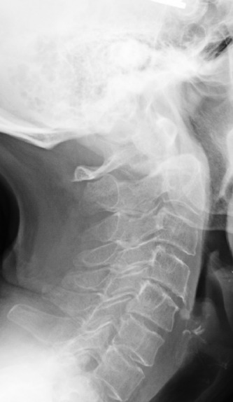 |
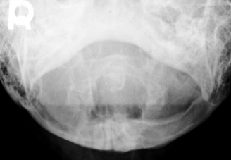
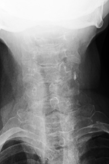
1. What is the key finding?
There are obvious degenerative changes of the cervical spine as well as osteopenia. The key finding however is the posterior subluxation of the atlas (C1) on the axis (C2). This is evident by the disruption of the spinolaminar line. The opacity with the sclerotic borders in the expected location of the dens represents the anterior arch of C1. Posterior to that you can make out the subtle appearance of a rounded opacity representing the dens, displaced posteriorly from the body of C2.
Diagnosis - Unstable Os Odontoideum
2. What is the etiology of the main finding - trauma, congenital, other?
An os odontoideum was generally considered to be a failure of fusion of the odontoid secondary growth center with the C2 body. It can be seen with increased frequency in patients with Down’s syndrome, Morquio syndrome, and multiple epiphyseal dysplasia. Recently, however, the etiology has become more controversial, with some postulating they arise from a fracture of the odontoid synchondrosis prior to its closure at the age of 5-6.
An os odontoideum may be confused with an acute fracture of the dens. An os may be differentiated from an acute fracture by its rounded sclerotic margins and wide gap between the bony fragments; a fracture will show a narrower gap with irregular non sclerotic margins.
The prevalence of os odontoideum is generally unknown and is usually detected incidentally or when patients become symptomatic. In cases where patients with an os odontoideum present with symptoms this may include local mechanical neck pain, headache, and torticollis, as well as neurologic symptoms including paresis, progressive myelopathy, weakness, and ataxia. Patients with an unstable os may present with neurovascular symptoms such as ataxia, syncope, vertigo, and visual disturbances due to vertebral artery compression.
An os odontoideum can be stable or unstable. Flexion and extension radiographs are recommended to confirm stability, except in cases of obvious neurologic symptoms. A previously stable os odontoideum may become unstable due to new trauma or gradual increasing laxity of the supporting ligamentous structures. As such serial stress radiographs should be performed to asses for an interval change. In cases of sudden development of neurologic or neurovascular symptoms MRI assessment should be performed. In cases of instability or severe neurologic or neurovascular manifestations surgical intervention in the form of fusion is recommended. Imaging features that favour the need for surgery include a posterior ADI space (measured from the posterior margin of the os to the anterior border of the C1 posterior arch) measuring less that 13 mm, greater than 5 mm translation of C1 on C2, and evidence of pathologic changes in the cord on MRI.
Complications other than progression from stability to instability are rare. Complications in surgical cases include risks commonly associated with surgery and anesthesia, the potential for intra-operative neural or vertebral artery injury during hardware placement, and post-surgical infection. In rare and extreme occasions sudden death due to cord compression may occur.